Imbalances in neuronal excitation and inhibition have been proposed as an overarching explanation of at least some features of autism spectrum disorder (ASD), although they have typically been measured in model systems carrying rare mutations associated with the condition. In addition to continued work on this topic in mice, a new study establishes biomarkers of E/I balance in humans that are derived from neuroimaging data, and links them to sex differences in ASD at the neural and behavioral levels.
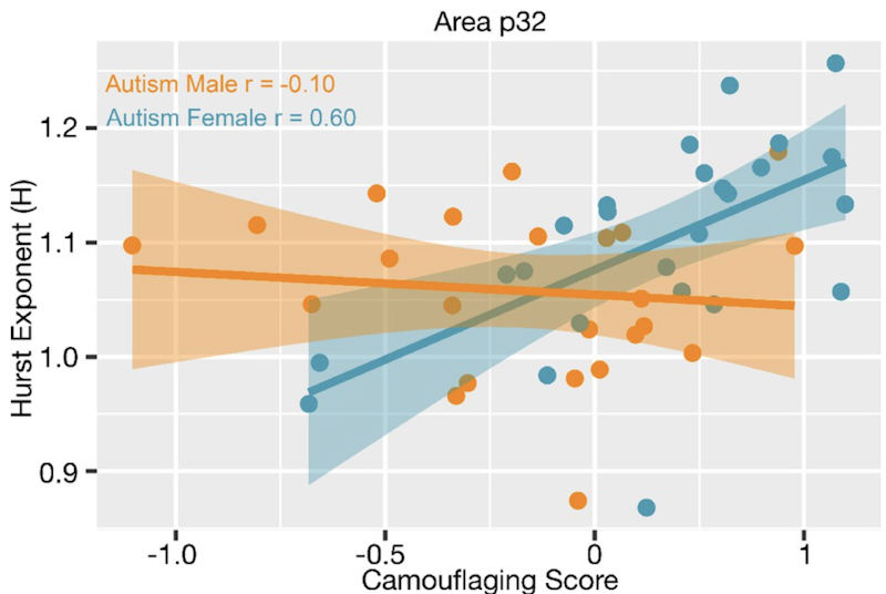
The work was supported in part by an Explorer Award to SFARI Investigator Stefano Panzeri, who collaborated with a group of investigators to develop new methods to measure E/I balance in human participants and to link it to other aspects of the condition. Specifically, Panzeri and colleagues used neuroimaging readouts such as local field potential and blood oxygenation levels to determine measures—including the Hurst exponent (H, related in some sense to neural noise)—that index E/I balance. They showed that H is reduced in the ventral medial prefrontal cortex in males with ASD, but not in females. Moreover, the authors reported an association between levels of H in females with ASD and their ability to compensate for, or camouflage, difficulties in social communication that define the disorder.
Of note, the investigators also reported that autism-associated genes that are known to affect neuronal excitation are highly overlapping with the set of genes whose expression is induced by androgens in neural stem cells. This result suggests that sex hormone-dependent gene expression may initiate a cascade of events leading to altered E/I balance. The ability to measure this imbalance using neuroimaging methods in individuals with idiopathic ASD should be a boon to research efforts aimed at understanding how the known sex bias arises.

Reference(s)
Intrinsic excitation-inhibition imbalance affects medial prefrontal cortex differently in autistic men versus women.
Trakoshis S., Martínez-Cañada P., Rocchi F., Canella C., You W., Chakrabarti B., Craig M.C., Daly E.M., Deoni S.C.L., Ecker C., Happé F., Henty J., Jezzard P., Johnston P., Jones D.K., Lai M.-C., Madden A., Mullins D., Murphy C.M., Murphy D., Pasco G., Ruigrok A.N., Sadek S.A., Spain D., Stewart R., Suckling J., Wheelwright S.J., Williams S.C., Baron-Cohen S., Gozzi A., Lai M.-C., Panzeri S., Lombardo M.


